Home \ Speciality Clinics


Speciality Clinics
Our Base hospital in Calicut has all the various Speciality Divisions, which are manned by well qualified and experienced Doctors/Para Medics.
- CATARACT AND COMPREHENSIVE OPHTHALMOLOGY
- RETINA CLINIC (SURGICAL AND MEDICAL)
- SURGICAL RETINA
- UVEITIS & OCULAR INFLAMMATION
- NEURO - OPHTHALMOLOGY
- PAEDIATRIC OPHTHALMOLOGY AND STRABISMUS (SQUINT)
- GLAUCOMA
- CORNEA CLINIC & OCULAR SURFACE
- CONTACT LENS CLINIC
- LASIK
- OCULOPLASTIC SURGERY
- ANESTHESIA
- PHARMACY
- LABORATORY
- OPTICAL STORE
Cataract and Comprehensive Ophthalmology
Small incision Phaco cataract surgeries are conducted by experienced surgeons. More than 10000 surgeries are done every year.
Main activities
- Foldable IOL
- Bifocal IOL
- Bifocal Foldable IOL
- Small Incision Phaco Surgery
- Phaco Trab
- Toric IOL
- Microincision Cataract Surgery (MICS)

Our Doctors


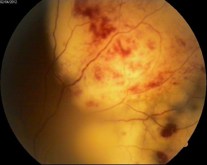
Retina Clinic (Surgical And Medical)
Main activities Medical Retina
1. Advanced center for ocular imaging
- Digital fundus photography.
- Optos wide field Photography.
- Mobile camera based telescreening.
- Fundus fluorescein angiography.
- Indocyanine angiography.
- Ultrasound B-scan.
- Ultrasound biomicroscopy.
- Laser indirect ophthalmoscopy.
- Spectral domain optical coherence tomography.
- Fundus autofluorescence imaging.

2. Retinal Electrophysiology
- ERG
- MFERG
- VEP
- EOG
3. Retinal laser Center
- Retinal laser for Diabetic retinopathy.
- ROP laser center for premature babies.
- Photo Dynamic therapy (PDT).
- Transpupillary thermal therapy (TTT) for Retinoblastoma.
- Micropulse Laser.

4. Intravitreal injection/Implants
- Intravitreal Anti VEGF drugs and steroid implants.
Our Doctors


Surgical Retina
Dedicated retina or with wide angle retinal viewing systems (BIOM & RESIGHT). Alcon constellation-micro incision vitrectomy surgery for complicated retinal disease like diabetic retinopathy, macular hole, retinal detachment, ocular trauma, intraocular foreign bodies, Intraocular tumours.
- Retinal detachment surgery
- Vitrectomy surgery for vitreous hemorrhage and other retinal diseases, diabetic eye diseases etc.
- Anti vegf Therapy
- Intra Ocular foreign body removal
- Ocular Trauma
- Macular hole surgery
Our Doctors
Uveitis & Ocular Inflammation
Main activities
- Treatment of ocular inflammation
- Uveitis
- Ocular infections/endophthalmitis
- Advanced diagnostics with Vitreous biopsy


Neuro - Ophthalmology
Main activities
- Optic neuritis
- Ischemic opticneuropathy
- Ocular myasthenia
- Hemifacial spasm
- Ocular Cranial Nerve Palsy

Paediatric Ophthalmology And Strabismus (Squint)
Main activities
The department of Paediatric Ophthalmology & Strabismus at Comtrust eye hospital features expert Clinical experiences and state of the art refraction and diagnostic services.
We realise with strong conviction that the benefits of timely detection and management of visual problems in childhood goes a long way in shaping a person's Career and future.
1.Binocular vision assessment & Management-
a) Medical
- Visual assessment & glass prescription.
- PRISM therapy
- Assessment & spectacle /prism management of Squint (Adult & paediatric).
- Amblyopia (Lazy eye) therapy including dichoptic Therapy (Bynocs).
- Assessment of Binocular vision anomaly & its management.
- Cerebral visual Impairment (CVI) screening.
- LOW Vision AID (LVA) provision to enhance those children with visual Impairment.
b) Surgical:
- Invasive and non-Invasive squint surgery. (Adult & Paediatric, under anaesthesia appropriate for age (TA/LA/GA).
- Surgery for Nystagmus. Including topical one stage adjustable surgery.
- Botulinum toxin (Botox) Injection.
2) Paediatric cataract surgery.
3) Paediatric glaucoma surgery (Goniotomy, Trabeculotomy, Trabeculectomy with MMC, Glaucoma Drainage Devices) and Examination under anaesthesia.
4) Paediatric Oculoplasty services
- Probing.
- Congenital and Acquired Adnexal Diseases.
- Traumatic lid & Canalicular Repair.
- Ptosis correction.
5) Pupilloplasty.
6) Paediatric ocular trauma Management.
7) Retinopathy of Prematurity (ROP) screening in collaboration with Retina department.
8) Paediatric orbital surgeries in collaboration with Oculoplasty department.
Instruments
- Hand held slit lamp
- Hand held keratometer (Retinomax).
- Ultrasound A scan (Sonomed).
- Open space Auto refractometer.
- Perkins hand held tonometer.
- Orthoptic Evaluation
- Kay picture fixation sticks & Lang fixation cube.
- Synaptophere.
- Loose prism set.
- Prism Bar.
- TNO /Lang Test.
- Worth 4 dot Test.
- Binocular Vision assessment
- RAF Rule.
- Accommodation flipper.
- Vergence Facility Prisms.
- Paediatric Vision Assessment.
- TA (Teller Acuity) cards
- Sheridan Gardiner test.
- Allen picture chart.
- Lea Symbols / Paddles.
- Log MAR Chart.
- Diplopia Light | Hess chart.
- Electrophysiological Tests: ERG/VEP.
- Bynocs - Amblyopia Therapy.
- Gaze Lab for Nystagmus.
- Optokinetic Drum. (OKN Drum).
- IOL Master 500 and 700.
- Topography/As OCT (Anterior Segment OCT).
- Ultrasound Biometry.
- Lasik for anisometropic amblyopia.
Publications and Research:
- Role of over minus therapy in Intermittent Exotropia.
- Surge in Acute Acquired Comitant Esotropia during Covid Lockdown.
- A Randomized controlled trial to evaluate the effect of low dose Atropine on the progression of childhood myopia in South India.
- Surgical Outcomes of graded inferior oblique recession with or without anteriorisation on over elevation in adduction.
- Etiology and associations of paediatric cataract and long-term visual outcomes following paediatric cataract surgery.
- Visual Outcomes following paediatric ocular trauma.
- Congenital ectropion uvea with glaucoma an early sign of neurofibromatosis 1.
- Brittle cornea Syndrome – A rare genetic disease.
- Anterior necrotizing scleritis? S/P covid a rare presentation
Our Doctors


Glaucoma
Glaucoma is a chronic, progressive eye disease caused by damage to the optic nerve, which leads to visual field loss. One of the major risk factors is eye pressure. An abnormality in the eye’s drainage system can cause fluid to build up, leading to excessive pressure that causes damage to the optic nerve. The optic nerve is a bundle of nerve fibers that connects the retina with the brain. This damage leads to loss of eyesight.
The vision loss starts out in the edges of the visual field and slowly impacts the central vision. It takes months to years after the nerve damage has occurred before you may notice the symptoms. Once vision is lost, it cannot be recovered.
Most people with glaucoma do not notice symptoms until they begin to lose eyesight. As glaucoma damages optic nerve fibers, small blind spots may begin to develop. These spots usually occur on the side or in the peripheral vision. Many people do not notice the blind spots until significant optic nerve damage has already happened. Blindness can result when the entire nerve is destroyed.
- Glaucoma is the leading cause of irreversible blindness.
- Glaucoma is known as the “silent blinder” because there are no noticeable symptoms in the early stages.
- Early detection and treatment for glaucoma are the most important steps to prevent vision loss.
- Family history, especially a sibling relationship, is a risk factor for glaucoma.
- Eye pressure is the only known modifiable risk factor for glaucoma and is the target of current treatment regimens.
There are many types of glaucoma:
- Open-angle glaucoma — The most common form of glaucoma, this type is caused by damage to the filter in the eye’s drainage canals.
- Angle-closure glaucoma — This type of glaucoma is caused by a rapid blockage of the eye’s drainage canals due to a closed or narrow angle between the iris and cornea where the filter is located.
- Low-tension or normal-tension glaucoma — A type of glaucoma in which damage occurs to the optic nerve without eye pressure exceeding its normal range.
- Congenital glaucoma — A type of glaucoma that occurs in infants when there are incorrect or underdeveloped drainage canals in the eye during the prenatal period.
- Uveitic (inflammatory) glaucoma — This type of glaucoma caused by autoimmune and inflammatory disorders.
- Neovascular glaucoma — A type of glaucoma associated with poorly controlled diabetes and other conditions that damage the blood vessels in the body.
- Race. Glaucoma is the leading cause of blindness for people of African descent.
- Age. People age 60 and older are more at risk for developing glaucoma.
- Family history. People with a family history of glaucoma are more likely to develop the disease, especially those with a sibling who has the condition.
- High fluid pressure inside the eyes. People with high fluid pressure inside the eyes are at an increased risk.
- Decreased corneal thickness. People with a thinner cornea are at greater risk of glaucoma
- Blurred or narrowed field of vision
- Severe pain in the eyes
- Seeing halos or “rainbows” around lights
- Nausea
- Vomiting
- Headache
- Visual acuity test. The common eye chart test measures how well you can see at various distances.
- Pupil dilation. The pupil is widened with eye drops to allow a close-up exam of the eye’s optic nerve and retina.
- Visual field. This test measures a person’s side or peripheral vision. Lost peripheral vision may mean a person has glaucoma.
- Tonometry. This standard test determines the fluid pressure inside the eye.
- Optic nerve imaging. Photographs of the optic nerve are taken to indicate areas of damage.
- Gonioscopy: A lens is placed on the eye to look at the area called the drainage angle. This is where fluid drains from the eye. This test determines whether the drain is open or closed, and if any damage has occurred.
- Pachymetry: A measurement is taken of corneal thickness.
- Central Corneal Thickness.
- Medicines. Some medicines cause the eye to make less fluid, while others lower pressure by helping fluid drain from the eye.
- Laser surgery. Several types of surgery using a laser are used to treat glaucoma.
- Surgery. The purpose of surgery is to create a new opening for fluid to leave the eye. This can be done by creating a passage for drainage or by implanting a shunt to help drain the fluid.
- Trabeculectomy
- Goniotomy
- Phacotrab.
Glaucoma Causes
Many factors lead to glaucoma. While increased eye pressure is the only known modifiable risk factor known at this time, is it not a cause. Glaucoma can also develop with normal eye pressure.
Glaucoma Risk Factors
Anyone can develop glaucoma. However, some people are at higher risk than others. Risk factors for glaucoma include:
Anyone in these risk groups should get an annual eye exam.
Glaucoma Symptoms
Most glaucoma sufferers do not experience symptoms until they start to lose their vision. Small blind spots may start to form as the optic nerve fibres are damaged by glaucoma. These spots typically appear in the side or peripheral vision. Many people do not become notice of the blind spots until there has been significant optic nerve damage. Blindness can result when the entire nerve is destroyed.
Symptoms of Acute-Closure Glaucoma
One type of angle-closure glaucoma, called acute angle-closure glaucoma, does produce noticeable symptoms. This is because there is a quick buildup of pressure in the eye. Each person may experience symptoms differently, which include:
The symptoms of acute angle-closure glaucoma may resemble those of other eye problems. Receiving medical attention quickly when first noticing symptoms can help prevent blindness.
Glaucoma Diagnosis
Your eye health care provider will take your complete medical history and examine your eyes. You may also have the following tests to diagnose glaucoma:
Glaucoma Treatment
Although there is no lasting treatment for glaucoma, early treatment can often control it. This may include:
In some cases, a single surgery isn't enough to slow down the progress of glaucoma. Repeat surgery and/or continued treatment with medicines may be necessary. Without treatment, glaucoma can cause permanent blindness
Our Doctors


Cornea Clinic & Ocular Surface
Main activities
- Pterygium surgery Pioneer in conjunctival autograft
- Corneal Transplantation
- Our own Eye Bank (approved by Eye Bank Association of India) is able to support our corneal transplantation
- C3R
- Corneal Transplantation
- Refractive Surgery
- Lasik
- Phakic IOL


Our Doctors


Contact Lens Clinic
This clinic dispenses all kinds of contact lenses under proper diagnosis and supervision of Specialist doctors.

Lasik
We offer premium corneal examination using VISANTE ANTRIOR OCT, ATLAS CORNEAL TOPOGRAPHY and customized treatment plan using CRS MASTER and Lasik treatment with our specialized equipment MEL 80 EXCIMER laser system.

Oculoplastic Surgery
Main activities
- Ptosis Surgery
- Lacrimal sac-dcr dct,fistulectomy, lacrimal intubation
- Ectropion, Entropion correction
- Blepharoplasty
- Socket reconstruction
- Botox
- Orbit tumor surgery
- Cosmetic Lid Surgery
Oculoplasty is a branch of ophthalmology which deals with the diseases of the structures surrounding the eye such as
- Eyelids
- Lacrimal system apparatus/ tear drainage problems
- Bony socket
- Orbital tissues
The specialised surgeries performed in such cases are referred to as ophthalmic plastic surgery/oculoplastic surgery.
Diseases affecting the eyelids are
- Infection – stye, chalazion
- Droopy lids (from birth/acquired) – (ptosis)
- Lid malpositions – outrolling (ectropion), inward rolling (entropion), incomplete closure of the eye (lagophthalmos), excessive opening of the eye(eyelid retraction)
- Lid tumours – benign and cancers of the lid
- Eyelash related problems such as misdirected lashes (trichiasis)
- Congenital abnormalities of lid – absent lids, partially formed lids (coloboma)
- Trauma (accident)– lid tears and lid defects following surgery for removal of lid cancers
- Age-related excessive skin over lids or lower lid bags, drooping of brows
Diseases affecting the tear drainage system
- Excessive watering since birth - congenital NLD block
- Inflammation of the lacrimal sac – adults (dacryocystitis)
- Watering in adults due to tear outflow problems
- Canalicular tears due to trauma
- Infective such as orbital cellulitis and collection of pus abscess
- Inflammatory conditions – inflammation of muscles surrounding the eye or lacrimal gland
- Tumours behind or around the eye benign and cancerous – both in children or adults
- Thyroid eye disease
- Congenital absence of eyeball/shrinking of the eyeball due to acquired causes
Diseases affecting the bony socket include orbital fractures following trauma Diseases affecting the orbit include
Diseases of the ocular surface – tumours/cancers (ocular surface squamous neoplasia) and inflammations
The treatment of these diseases are either medical or surgical, managed by a specialised Oculoplasty Consultant. The surgeries performed are planned such that the root cause of the disease is addressed. Various specialised surgeries performed here are ptosis surgeries, lid malposition surgeries, cosmetic lid surgeries, lid tear repairs/lid reconstruction, lacrimal procedures – probing under endoscopic guidance, dacryocystectomy(DCT), dacryocystorhinostomy(DCR), lacrimal intubation, orbitotomies for tumour removal, orbital fracture repair, corrective surgeries for thyroid eye disease, removal of the eye and implant surgeries for cosmetic rehabilitation, botulinum toxin injections for involuntary facial movements and cosmetic indications. Surgeries are usually day-care with admissions only in exceptional cases needing monitoring.
We have a prosthesis department for cosmetic rehabilitation of patients who have an absent eye since birth or following surgery or accident. Customised ocular prosthesis is dispensed for each patient with simple instructions of use by our ocularistOur Doctors

Anesthesia
Since its inception in 1999, The Department of Anaesthesiology and Critical care serve its patients as independent department at Comtrust Charitable Trust Eye Hospital providing exemplary, compassionate patient care through a consistent commitment to evidence-based, patient-centered medicine. The Department of Anaesthesiology and Critical care staff members are a diverse, highly integrated and collaborative group of adults and pediatric anesthesiologists, assisted by trained and qualified anaesthesia technologists committed to provide the best possible care to the patients. The Department aims to pursue research that will define novel therapies in a dynamic academic environment and advance the standards for delivery of excellent patient care through the development of innovative application of processes and technology. The Department of Anaesthesiology and Critical care offers outstanding anaesthetic and critical care for the entire Comtrust Eye Hospitals Patients. Our anaesthesiologists care for more than 20,000 people each year. This large volume, integrated with ongoing research and dynamic education, results in wide-ranging expertise in the anaesthetic and perioperative management of patients.
We have 8 anaesthesiologits, including the visiting specialist consultants, assisted by anaesthesia technologists, post op nurses and intensivist, providing care in a wide variety of settings including the operating rooms, intensive care units, out-station anaesthesia, and pain medicine in the hospital and affiliated centres under Comtrust Charitable Trust Eye hospital. The Department coordinates and conducts BLS, ACLS training to the entire staffs of the hospital. While many members of the department conduct impactful basic and clinical research, many others teach learner-forward postgraduates, anaesthesia technologits and continuing education courses. The Department offers training courses for the anaesthesia doctors and technologists.
Our Doctors


Pharmacy
Hospital has a well-stocked Pharmacy which caters for all types of eye care related medicines
Laboratory
- A fully equipped laboratory is functioning at the hospital.
- Heamotology Analyser.
- Detection of Infectious disease in Biochemistry department.
- Genru- PA 50 machine which is used to detect HbA1c levels in Diabetic Retinopathy patients.
- Clinical pathology tests deals with the diagnosis of diseases usually based on laboratory analysis of blood or urine.
Optical Store
Hospital has a Optical Retail Outlet where good quality optical frames/lenses/other products are available at fair prices. This shop has a large stock of quality products (both imported and Indian) which are offered as per correct specifications prescribed by doctors.